Chaukhambha Orientalia Chest X-Ray and Its Interpretation
Chaukhambha Orientalia Chest X-Ray and Its Interpretation
Couldn't load pickup availability
Share
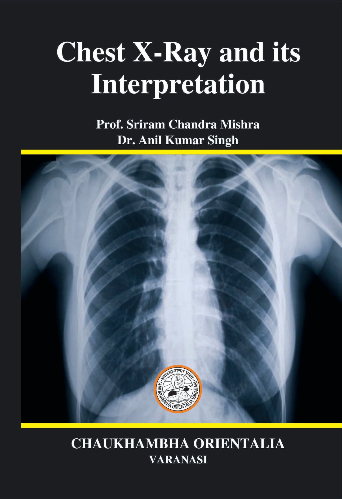
Chaukhambha Orientalia Chest X-Ray is a radiological imaging technique used to visualize the structures within the chest cavity, including the lungs, heart, and surrounding tissues. The interpretation of a chest X-ray involves analyzing the various structures and looking for any abnormalities that may indicate underlying medical conditions.
Here is a detailed description of the key components of a Chaukhambha Orientalia Chest X-Ray and their interpretation:
1. **Lungs**: The lungs appear as dark areas on the X-ray due to their air-filled nature. Abnormalities in the lungs, such as infiltrates (areas of increased density), nodules, masses, or consolidation (solidification of lung tissue), may indicate conditions such as pneumonia, lung cancer, or pulmonary edema.
2. **Heart**: The heart is typically seen as a well-defined shadow in the center of the chest. Enlargement of the heart, indicated by an increased cardiothoracic ratio, may suggest conditions such as heart failure or cardiomegaly.
3. **Diaphragm**: The diaphragm is a dome-shaped muscle that separates the chest cavity from the abdominal cavity. Abnormalities in the diaphragm, such as elevation or flattening, may be seen in conditions such as diaphragmatic paralysis or hernias.
4. **Trachea and Bronchi**: The trachea (windpipe) and bronchi (airways leading to the lungs) are visible as tubular structures on the X-ray. Deviation of the trachea or bronchial narrowing may indicate conditions such as tracheal deviation or bronchial obstruction.
5. **Bones and Soft Tissues**: The chest X-ray may also show the ribs, clavicles, and soft tissues of the chest wall. Fractures, bony lesions, or soft tissue abnormalities may be identified on the X-ray.
6. **Mediastinum**: The mediastinum is the central compartment of the chest that contains the heart, great vessels, esophagus, and other structures. Abnormalities in the mediastinum, such as mediastinal widening or masses, may indicate conditions such as mediastinitis or mediastinal tumors.
7. **Pleura**: The pleura is a thin membrane that lines the chest cavity and covers the lungs. Pleural effusions (accumulation of fluid in the pleural space) or pleural thickening may be visible on the X-ray and can indicate conditions such as pleurisy or pleural tumors.
Overall, the interpretation of a Chaukhambha Orientalia Chest X-Ray involves a systematic evaluation of the various structures within the chest cavity to identify any abnormalities that may help in diagnosing and managing a wide range of respiratory, cardiac, and other medical conditions. It is important to consult with a radiologist or healthcare provider for a comprehensive assessment and appropriate management based on the findings of the chest X-ray.